Section 24-1 Review Viral Structure and Replication Answers
Chapter 12: Introduction to the Immune System and Disease
12.one Viruses
Learning Objectives
By the end of this section, you will exist able to:
- Depict how viruses were kickoff discovered and how they are detected
- Explain the detailed steps of viral replication
- Draw how vaccines are used in prevention and handling of viral diseases

No 1 knows exactly when viruses emerged or from where they came, since viruses do not exit historical footprints such as fossils. Mod viruses are idea to be a mosaic of bits and pieces of nucleic acids picked up from various sources forth their respective evolutionary paths. Viruses are acellular, parasitic entities that are not classified within any domain because they are non considered alive. They accept no plasma membrane, internal organelles, or metabolic processes, and they do non divide. Instead, they infect a host cell and apply the host's replication processes to produce progeny virus particles. Viruses infect all forms of organisms including leaner, archaea, fungi, plants, and animals. Living things grow, metabolize, and reproduce. Viruses replicate, but to do and so, they are entirely dependent on their host cells. They do not metabolize or grow, just are assembled in their mature class.
Viruses are various. They vary in their structure, their replication methods, and in their target hosts or even host cells. While virtually biological diversity tin can exist understood through evolutionary history, such every bit how species have adjusted to conditions and environments, much near virus origins and evolution remains unknown.
How Viruses Replicate
Viruses were kickoff discovered afterward the evolution of a porcelain filter, called the Chamberland-Pasteur filter, which could remove all bacteria visible nether the microscope from whatever liquid sample. In 1886, Adolph Meyer demonstrated that a disease of tobacco plants, tobacco mosaic disease, could exist transferred from a diseased establish to a healthy ane through liquid establish extracts. In 1892, Dmitri Ivanowski showed that this affliction could be transmitted in this way even after the Chamberland-Pasteur filter had removed all viable bacteria from the extract. Still, it was many years earlier it was proven that these "filterable" infectious agents were not simply very modest leaner but were a new type of tiny, affliction-causing particle.
Virions, single virus particles, are very small, about 20–250 nanometers (1 nanometer = 1/1,000,000 mm). These individual virus particles are the infectious grade of a virus outside the host cell. Dissimilar leaner (which are about 100 times larger), we cannot see viruses with a calorie-free microscope, with the exception of some large virions of the poxvirus family (Figure 12.three).
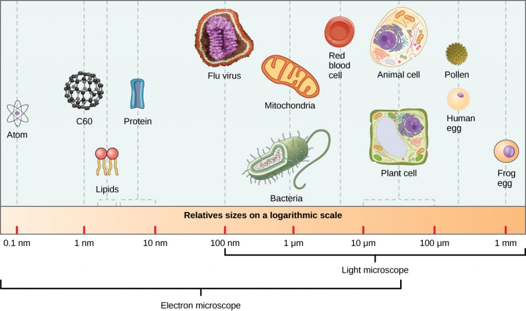
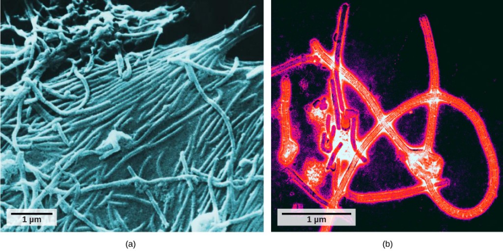
Information technology was not until the development of the electron microscope in the 1940s that scientists got their first good view of the structure of the tobacco mosaic virus (Figure 12.2) and others. The surface structure of virions can be observed by both scanning and manual electron microscopy, whereas the internal structures of the virus can only exist observed in images from a transmission electron microscope (Figure 12.four).
The use of this applied science has allowed for the discovery of many viruses of all types of living organisms. They were initially grouped by shared morphology, meaning their size, shape, and distinguishing structures. Later, groups of viruses were classified by the blazon of nucleic acrid they contained, Dna or RNA, and whether their nucleic acid was single- or double-stranded. More recently, molecular analysis of viral replication cycles has further refined their classification.
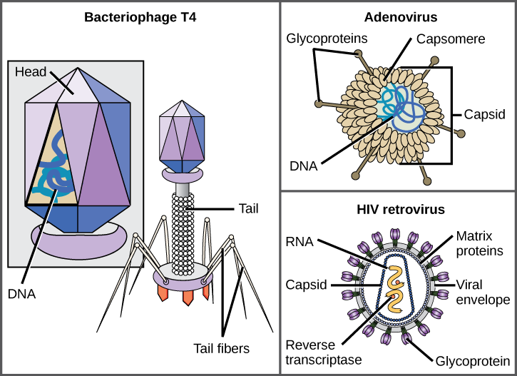
A virion consists of a nucleic-acid core, an outer protein coating, and sometimes an outer envelope made of poly peptide and phospholipid membranes derived from the host cell. The most visible difference between members of viral families is their morphology, which is quite diverse. An interesting feature of viral complexity is that the complication of the host does not correlate to the complexity of the virion. Some of the most complex virion structures are observed in bacteriophages, viruses that infect the simplest living organisms, bacteria.
Viruses come in many shapes and sizes, but these are consistent and distinct for each viral family unit (Effigy 12.5). All virions have a nucleic-acid genome covered past a protective layer of poly peptide, chosen a capsid. The capsid is fabricated of protein subunits chosen capsomeres. Some viral capsids are simple polyhedral "spheres," whereas others are quite complex in construction. The outer structure surrounding the capsid of some viruses is called the viral envelope. All viruses apply some sort of glycoprotein to attach to their host cells at molecules on the cell called viral receptors. The virus exploits these cell-surface molecules, which the cell uses for some other purpose, every bit a manner to recognize and infect specific cell types. For example, the measles virus uses a cell-surface glycoprotein in humans that normally functions in immune reactions and peradventure in the sperm-egg interaction at fertilization. Zipper is a requirement for viruses to afterward penetrate the cell membrane, inject the viral genome, and complete their replication inside the jail cell.
The T4 bacteriophage, which infects the Due east. coli bacterium, is among the nearly complex virion known; T4 has a protein tail structure that the virus uses to attach to the host prison cell and a head structure that houses its Deoxyribonucleic acid.
Adenovirus, a nonenveloped animate being virus that causes respiratory illnesses in humans, uses protein spikes protruding from its capsomeres to attach to the host prison cell. Nonenveloped viruses likewise include those that cause polio (poliovirus), plantar warts (papillomavirus), and hepatitis A (hepatitis A virus). Nonenveloped viruses tend to be more robust and more than probable to survive under harsh conditions, such as the gut.
Enveloped virions similar HIV (human immunodeficiency virus), the causative agent in AIDS (acquired allowed deficiency syndrome), consist of nucleic acid (RNA in the case of HIV) and capsid proteins surrounded by a phospholipid bilayer envelope and its associated proteins (Figure 12.5). Chicken pox, influenza, and mumps are examples of diseases acquired by viruses with envelopes. Because of the fragility of the envelope, nonenveloped viruses are more than resistant to changes in temperature, pH, and some disinfectants than enveloped viruses.
Overall, the shape of the virion and the presence or absence of an envelope tells us trivial most what diseases the viruses may cause or what species they might infect, just is withal a useful ways to begin viral classification.
Which of the post-obit statements about virus construction is true?
A) All viruses are encased in a viral membrane.
B) The capsomere is made up of small poly peptide subunits called capsids.
C) Dna is the genetic material in all viruses.
D) Glycoproteins help the virus attach to the host cell.
<!–D–>
Unlike all living organisms that use DNA as their genetic material, viruses may use either DNA or RNA as theirs. The virus core contains the genome or total genetic content of the virus. Viral genomes tend to be small compared to bacteria or eukaryotes, containing simply those genes that code for proteins the virus cannot go from the host cell. This genetic textile may exist single-stranded or double-stranded. It may as well be linear or round. While most viruses contain a single segment of nucleic acid, others have genomes that consist of several segments.
DNA viruses have a Deoxyribonucleic acid core. The viral DNA directs the host cell'southward replication proteins to synthesize new copies of the viral genome and to transcribe and interpret that genome into viral proteins. Dna viruses cause human diseases such equally chickenpox, hepatitis B, and some crabs diseases like canker and genital warts.
RNA viruses contain but RNA in their cores. To replicate their genomes in the host prison cell, the genomes of RNA viruses encode enzymes non found in host cells. RNA polymerase enzymes are not equally stable as Deoxyribonucleic acid polymerases and oft make mistakes during transcription. For this reason, mutations, changes in the nucleotide sequence, in RNA viruses occur more frequently than in DNA viruses. This leads to more rapid evolution and modify in RNA viruses. For example, the fact that influenza is an RNA virus is one reason a new influenza vaccine is needed every yr. Human diseases caused by RNA viruses include hepatitis C, measles, and rabies.
Viruses tin be seen as obligate intracellular parasites. The virus must adhere to a living cell, be taken inside, industry its proteins and copy its genome, and detect a fashion to escape the cell so the virus can infect other cells and ultimately other individuals. Viruses can infect merely sure species of hosts and only certain cells within that host. The molecular basis for this specificity is that a particular surface molecule, known every bit the viral receptor, must exist found on the host cell surface for the virus to attach. Also, metabolic differences seen in different cell types based on differential gene expression are a likely factor in which cells a virus may use to replicate. The cell must be making the substances the virus needs, such every bit enzymes the virus genome itself does not accept genes for, or the virus will not be able to replicate using that cell.
Steps of Virus Infections
A virus must "take over" a jail cell to replicate. The viral replication cycle can produce dramatic biochemical and structural changes in the host jail cell, which may cause cell damage. These changes, called cytopathic effects, tin change prison cell functions or even destroy the cell. Some infected cells, such every bit those infected by the mutual common cold virus (rhinovirus), die through lysis (bursting) or apoptosis (programmed cell expiry or "jail cell suicide"), releasing all the progeny virions at once. The symptoms of viral diseases consequence from the allowed response to the virus, which attempts to control and eliminate the virus from the body, and from prison cell impairment caused past the virus. Many animal viruses, such equally HIV (man immunodeficiency virus), leave the infected cells of the immune organisation by a process known as budding, where virions leave the jail cell individually. During the budding process, the prison cell does non undergo lysis and is non immediately killed. However, the impairment to the cells that HIV infects may get in impossible for the cells to part as mediators of amnesty, even though the cells remain live for a menstruation of fourth dimension. Most productive viral infections follow similar steps in the virus replication bike: attachment, penetration, uncoating, replication, assembly, and release.
A virus attaches to a specific receptor site on the host-cell membrane through zipper proteins in the capsid or proteins embedded in its envelope. The zipper is specific, and typically a virus volition only attach to cells of one or a few species and merely sure cell types inside those species with the advisable receptors.
Concept in Action

View this video for a visual explanation of how HIV and influenza attack the torso.
Dissimilar animal viruses, the nucleic acid of bacteriophages is injected into the host prison cell naked, leaving the capsid outside the cell. Plant and beast viruses can enter their cells through endocytosis, in which the cell membrane surrounds and engulfs the entire virus. Some enveloped viruses enter the cell when the viral envelope fuses directly with the cell membrane. Once inside the cell, the viral capsid is degraded and the viral nucleic acid is released, which then becomes available for replication and transcription.
The replication mechanism depends on the viral genome. Dna viruses normally use host cell proteins and enzymes to make additional Deoxyribonucleic acid that is used to copy the genome or be transcribed to messenger RNA (mRNA), which is then used in protein synthesis. RNA viruses, such as the influenza virus, unremarkably utilize the RNA core every bit a template for synthesis of viral genomic RNA and mRNA. The viral mRNA is translated into viral enzymes and capsid proteins to assemble new virions (Figure 12.6). Of form, in that location are exceptions to this pattern. If a host prison cell does non provide the enzymes necessary for viral replication, viral genes supply the information to direct synthesis of the missing proteins. Retroviruses, such every bit HIV, have an RNA genome that must be opposite transcribed to make DNA, which then is inserted into the host's DNA. To convert RNA into DNA, retroviruses contain genes that encode the virus-specific enzyme opposite transcriptase that transcribes an RNA template to DNA. The fact that HIV produces some of its own enzymes, which are not found in the host, has allowed researchers to develop drugs that inhibit these enzymes. These drugs, including the reverse transcriptase inhibitor AZT, inhibit HIV replication by reducing the activity of the enzyme without affecting the host's metabolism.
The last stage of viral replication is the release of the new virions into the host organism, where they are able to infect adjacent cells and repeat the replication bike. Some viruses are released when the host cell dies and other viruses can leave infected cells past budding through the membrane without directly killing the prison cell.
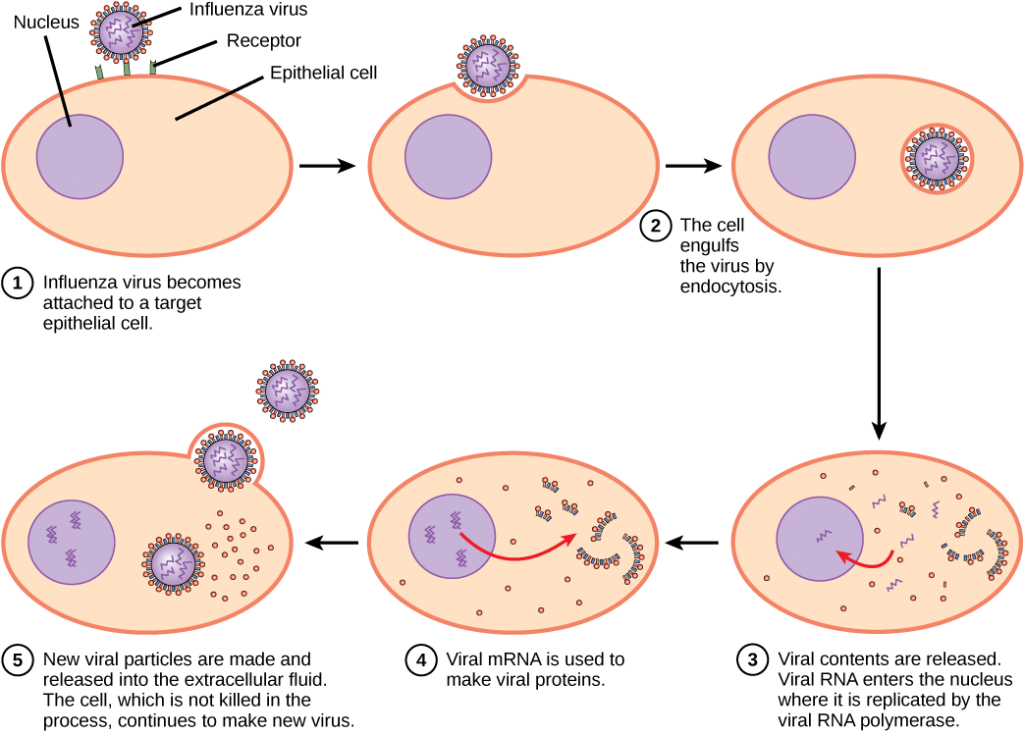
Flu virus is packaged in a viral envelope, which fuses with the plasma membrane. This way, the virus tin can get out the host cell without killing it. What advantage does the virus gain by keeping the host prison cell alive?
<!–The host cell can continue to make new virus particles.–>
Concept in Action

Click through this tutorial on viruses to identify structures, modes of manual, replication, and more.
Viruses and Affliction
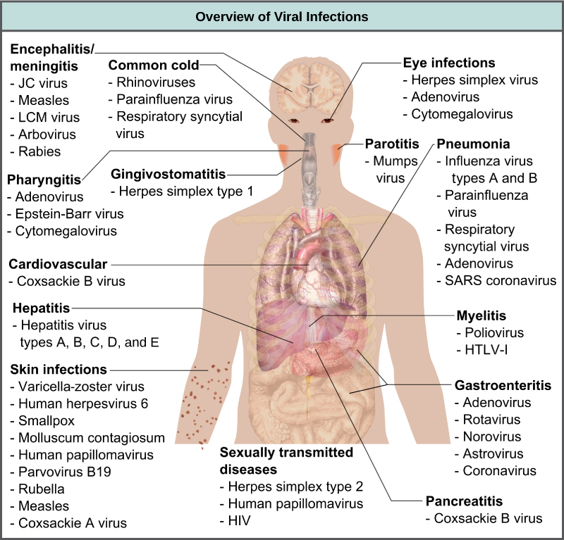
Viruses cause a diverseness of diseases in animals, including humans, ranging from the common cold to potentially fatal illnesses similar meningitis (Effigy 12.vii). These diseases can exist treated by antiviral drugs or by vaccines, but some viruses, such as HIV, are capable of avoiding the immune response and mutating so as to get resistant to antiviral drugs.
Vaccines for Prevention
While nosotros do take limited numbers of effective antiviral drugs, such equally those used to treat HIV and influenza, the chief method of decision-making viral disease is past vaccination, which is intended to prevent outbreaks by edifice immunity to a virus or virus family. A vaccine may be prepared using weakened live viruses, killed viruses, or molecular subunits of the virus. In full general, live viruses pb to meliorate immunity, but have the possibility of causing affliction at some low frequency. Killed viral vaccine and the subunit viruses are both incapable of causing disease, but in general atomic number 82 to less constructive or long-lasting immunity.
Weakened alive viral vaccines are designed in the laboratory to crusade few symptoms in recipients while giving them immunity against futurity infections. Polio was i disease that represented a milestone in the use of vaccines. Mass immunization campaigns in the U.S. in the 1950s (killed vaccine) and 1960s (live vaccine) essentially eradicated the disease, which caused muscle paralysis in children and generated fear in the general population when regional epidemics occurred. The success of the polio vaccine paved the manner for the routine impunity of babyhood vaccines against measles, mumps, rubella, chickenpox, and other diseases.
Alive vaccines are ordinarily made by attenuation (weakening) of the "wild-type" (disease-causing) virus by growing it in the laboratory in tissues or at temperatures dissimilar from what the virus is accepted to in the host. For example, the virus may exist grown in cells in a test tube, in bird embryos, or in alive animals. The adaptation to these new cells or temperature induces mutations in the virus' genomes, allowing them to grow better in the laboratory while inhibiting their ability to cause illness when reintroduced into the weather found in the host. These attenuated viruses thus nonetheless cause an infection, merely they do not grow very well, allowing the allowed response to develop in time to prevent major disease. The danger of using live vaccines, which are ordinarily more effective than killed vaccines, is the low simply significant risk that these viruses volition revert dorsum to their affliction-causing form by back mutations. Back mutations occur when the vaccine undergoes mutations in the host such that it readapts to the host and can again cause disease, which can then be spread to other humans in an epidemic. This happened as recently as 2007 in Nigeria where mutations in a polio vaccine led to an epidemic of polio in that country.
Some vaccines are in continuous development because sure viruses, such equally influenza and HIV, accept a high mutation rate compared to other viruses or host cells. With flu, mutation in genes for the surface molecules helps the virus evade the protective immunity that may accept been obtained in a previous influenza season, making it necessary for individuals to get vaccinated every year. Other viruses, such as those that cause the childhood diseases measles, mumps, and rubella, mutate so lilliputian that the aforementioned vaccine is used yr after yr.
Vaccines and Antiviral Drugs for Treatment
In some cases, vaccines can be used to treat an active viral infection. In the case of rabies, a fatal neurological affliction transmitted in the saliva of rabies virus-infected animals, the progression of the disease from the time of the animal seize with teeth to the time it enters the central nervous arrangement may exist ii weeks or longer. This is enough time to vaccinate an individual who suspects existence bitten past a rabid animal, and the boosted immune response from the vaccination is plenty to forbid the virus from entering nervous tissue. Thus, the fatal neurological consequences of the disease are averted and the private only has to recover from the infected seize with teeth. This arroyo is likewise existence used for the treatment of Ebola, one of the fastest and most deadly viruses affecting humans, though usually infecting limited populations. Ebola is also a leading crusade of death in gorillas. Transmitted past bats and bang-up apes, this virus can cause death in 70–90 per centum of the infected within two weeks. Using newly developed vaccines that heave the immune response, there is hope that immune systems of affected individuals will be better able to control the virus, potentially reducing mortality rates.
Another style of treating viral infections is the use of antiviral drugs. These drugs frequently have limited power to cure viral illness only have been used to control and reduce symptoms for a wide multifariousness of viral diseases. For near viruses, these drugs inhibit the virus by blocking the actions of one or more of its proteins. It is important that the targeted proteins be encoded for by viral genes and that these molecules are not present in a healthy host prison cell. In this way, viral growth is inhibited without damaging the host. In that location are big numbers of antiviral drugs bachelor to care for infections, some specific for a particular virus and others that tin can impact multiple viruses.
Antivirals have been developed to treat genital herpes (herpes simplex II) and flu. For genital canker, drugs such every bit acyclovir can reduce the number and elapsing of the episodes of active viral disease during which patients develop viral lesions in their skins cells. As the virus remains latent in nervous tissue of the body for life, this drug is not a cure but can brand the symptoms of the illness more manageable. For flu, drugs like Tamiflu can reduce the duration of "flu" symptoms by one or ii days, but the drug does not prevent symptoms entirely. Other antiviral drugs, such equally Ribavirin, have been used to treat a variety of viral infections.
By far the most successful use of antivirals has been in the handling of the retrovirus HIV, which causes a affliction that, if untreated, is ordinarily fatal within 10–12 years after being infected. Anti-HIV drugs take been able to control viral replication to the indicate that individuals receiving these drugs survive for a significantly longer time than the untreated.
Anti-HIV drugs inhibit viral replication at many different phases of the HIV replicative cycle. Drugs have been developed that inhibit the fusion of the HIV viral envelope with the plasma membrane of the host cell (fusion inhibitors), the conversion of its RNA genome to double-stranded DNA (reverse transcriptase inhibitors), the integration of the viral DNA into the host genome (integrase inhibitors), and the processing of viral proteins (protease inhibitors).
When whatsoever of these drugs are used individually, the virus' loftier mutation rate allows the virus to rapidly evolve resistance to the drug. The quantum in the treatment of HIV was the development of highly active anti-retroviral therapy (HAART), which involves a mixture of dissimilar drugs, sometimes called a drug "cocktail." By attacking the virus at unlike stages of its replication cycle, it is difficult for the virus to develop resistance to multiple drugs at the same fourth dimension. Still, even with the use of combination HAART therapy, there is concern that, over fourth dimension, the virus will evolve resistance to this therapy. Thus, new anti-HIV drugs are constantly beingness developed with the hope of continuing the boxing against this highly fatal virus.
Section Summary
Viruses are acellular entities that tin can usually just be seen with an electron microscope. Their genomes contain either Dna or RNA, and they replicate using the replication proteins of a host cell. Viruses are diverse, infecting archaea, bacteria, fungi, plants, and animals. Viruses consist of a nucleic-acid core surrounded by a protein capsid with or without an outer lipid envelope.
Viral replication inside a living cell always produces changes in the cell, sometimes resulting in cell death and sometimes slowly killing the infected cells. There are half dozen basic stages in the virus replication cycle: attachment, penetration, uncoating, replication, assembly, and release. A viral infection may exist productive, resulting in new virions, or nonproductive, meaning the virus remains inside the cell without producing new virions.
Viruses crusade a variety of diseases in humans. Many of these diseases can exist prevented by the use of viral vaccines, which stimulate protective immunity against the virus without causing major disease. Viral vaccines may also be used in active viral infections, boosting the power of the immune system to control or destroy the virus. Antiviral drugs that target enzymes and other protein products of viral genes accept been developed and used with mixed success. Combinations of anti-HIV drugs have been used to effectively control the virus, extending the lifespan of infected individuals.
Glossary
acellular: lacking cells
apoptosis: the prison cell death caused by induction of a cell's own internal mechanisms either every bit a natural pace in the development of a multicellular organism or by other environmental factors such as signals from cells of the allowed system
attenuation: the weakening of a virus during vaccine development
capsid: the protein coating of the viral core
cytopathic: causing cell damage
glycoprotein: a poly peptide molecule with attached carbohydrate molecules
vaccine: a weakened solution of virus components, viruses, or other agents that produce an immune response
virion: an private virus particle outside a host jail cell
viral envelope: a lipid bilayer that envelops some viruses
Source: https://opentextbc.ca/biology/chapter/12-1-viruses/